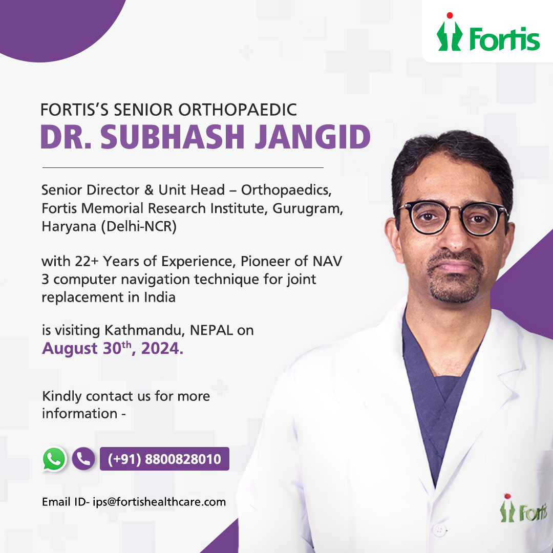
We are excited to announce that Fortis Hospital's esteemed medical expert, Dr. Subhash Jangid, Senior Director & Unit Head – Orthopaedics , Fortis Memorial Research Institute, Gurugram, will be in Nepal on 30th August 2024. Kindly contact us for more information, Contact/WhatsApp - +918800828010
About the Centre: Fortis Hospital, is one of the leading centers for advanced and comprehensive bone & joint care in the India and does the highest number of joint replacements in the region. With our legacy of clinical expertise, innovative technology and unparalleled success rates, a Centre of Excellence offers holistic care for Bone & Joint Problems. Our treatments involve minimally invasive surgeries for trauma recovery or even bone restructuring. Our team of anesthesiologists, rheumatologists and rehabilitation experts are here to make your recovery smooth and rapid. The hospital is equipped to perform an array of surgeries including uni-compartmental knee replacement, surface replacement of hip, arthroscopic management of knee, shoulder and ankle, and complex revision joint replacement surgeries. The hospital also provides state-of-the-art treatment for sports injuries, an osteoporosis clinic, and ankle and foot management. We have one of the very units in the region to house the most advanced computerized navigation system for joint replacement surgeries.
JOINT REPLACEMENT | ARTHRITIS | SPORTS MEDICINE | ARTHROSCOPY | COMPLEX TRAUMA | HIGH RISK CASES | SMALL JOINTS (HAND/WRIST/FOOT) | BONE CANCER| Foot and Ankle Expert

Why Choose Us
-
1. Experienced doctors backed by highly skilled paramedics
-
2. More than 22 years of clinical experience
-
3. Best in class medical services
-
4. State of the art medical technology
-
5. Best Sports Medicine and Joint replacement Doctors
-
6. Best Foot and ankle Expert
Our Team of Experts
Conditions and Treatments
- Treatment
-

Sports Medicine: Boon for Athletes
Excellence is not an act, but a habit. This holds true for any sportsperson as well as a sports injury specialist because there is a very narrow margin of error in the performance of both.
Sports Medicine specialty deals with diagnosis and treatment of sports injuries. Although the specialty gained limelight in recent years, sports physicians have existed from ancient times. Galen (131 to 201 AD), a famous ancient physician was a team doctor for gladiators in the Pegamum Kingdom. A sports medicine doctor is trained in treating musculoskeletal injuries.
TYPES OF INJURIES
Sports injuries primarily include ligament tears (ACL/PCL tears), fractures, recurrent dislocations including shoulder dislocation, rotator cuff injury, tennis elbow, pulled muscles, sprains and strains, Achilles Tendon injuries, frozen shoulder, shoulder impingement, hand injuries, mallet finger, trigger finger and trigger thumb. Almost all these injuries prevent the player from performing in professional and recreational sports.
WHY A SPORTS INJURY PHYSICIAN?
Delay in diagnosis and treatment of sports injuries always affects an athlete’s performance and career. A sports physician is specifically trained and equipped with the latest technology, expertise, knowledge and skills for treating such injuries. The sports physician will opt for a medical procedure which is important for faster and quicker recovery.
Sometimes, there are two options to treat a particular injury – one is non-operative but requires more time for recovery, while the other is operative, but the healing process is faster. The sports physician will always choose the second one and that, too, with minimal invasive surgical methods, so that morbidity and surgical scars are negligible.
MEDICAL & SURGICAL PROCEDURES
Sportspersons are offered a procedure which gives swift treatment and faster recovery. For this, a hospital should be well-equipped with specialised sports injury doctors and provide the latest technology such as MRI, CT scan, ultrasound etc. The various surgical procedures performed for sports injuries include:
Anterior Cruciate Ligament (ACL)
Arthroscopy of knees, shoulders and ankles
Cartilage restoration
Fracture repair (surgical and non-surgical)
Rotator cuff repair
Shoulder instability surgery
Tendon repair
Platelet rich plasma therapy
Osteochondritis dessicans - mainly for Osteoarthritis
OCD Talus & knee
Acromioclavicular joint
Ankle arthroscopy
Carpal tunnel syndrome release
PIP joint dislocation
Dupuytren's contracture -

Under Total Knee Replacement Surgery, the parts of the bones that rub together are resurfaced with metal and plastic implants. Using special, precision instruments, the damaged surfaces of the bones are removed and replacement surfaces are fixed into place. The surface of the femur is replaced with a rounded metal component that comes very close to matching the curve of your natural bone. The surface of the tibia/leg bone is replaced with a smooth plastic component. This flat metal component holds a smooth plastic piece made of ultra-high-molecular weight polyethylene
Q: How do I know if I need a Knee Replacement?
A. If you have difficulty walking or performing everyday activities, it may be time to consider Knee Replacement surgery. Doctors generally try to delay Total Knee Replacement for as long as possible in favor of less invasive treatments. However, for patients with advanced joint disease, knee replacement offers the chance for relief from pain and a return to normal activities plastic that serves as the cartilage. The undersurface of the kneecap may also be replaced with an implant made of the same polyethylene plastic.
Q. What happens during Knee Replacement Surgery?
A. On the day of the surgery, a small tube (intravenous line) will be inserted into your arm. This tube will be used to administer antibiotics and other medication during the surgery. You will then be taken to the operating room and given anesthesia. After the anesthesia takes effect, your knee will be scrubbed and sterilized with a special solution. The surgery will begin with an incision over the knee that will expose the joint. When the bones are fully visible to the surgeon, special, precision guides and instruments are used to remove the damaged surfaces and shape the ends of the bones to accept the implants. The implants are then secured to the bones. When the surgeon is satisfied with the fit and function of the implants, the incision will be closed.
A special drain may be inserted into the wound to drain the fluids that naturally develop at the surgical site. A sterile bandage will then be applied, and you will be taken to the recovery room, where you will be closely monitored. Your surgery will likely take between one and three hours, depending on individual circumstances. As your anesthesia wears off, you will slowly regain consciousness. A nurse will be with you, and may encourage you to cough or breathe deeply to help clear your lungs. You will also be given pain medication. When you are fully awake, you will be taken to your hospital room. Your knee will remain swollen and tender for a few days.
Q. How soon can I return to normal activities after surgery?
A. Within six weeks after surgery, most patients are able to walk with the help of a cane. You will probably feel well enough to drive within seven to eight weeks after surgery. In most cases, successful Joint Replacement Surgery will relieve your pain and stiffness, and allow you to resume many of your normal daily activities. But even after you have fully recovered from your surgery, you will still have some restrictions. Normal daily activities do not include contact sports or activities that put excessive strain on your joints. Although your artificial joint can be replaced, a second implant is seldom as effective as the first.
Q. What is Uni Condylar Replacement?
A. Uni Condylar Replacement also known as partial knee replacement is a very effective surgical treatment of for osteoarthritis with damaged confined to only one compartment of the knee joint. -

The aim of total hip replacement is to replace the head of the femur {ball} and reline the acetabulum {socket/cup} with man-made components. There are various types of hip replacements and your surgeon would have discussed with you the type of prosthesis you are likely to receive.
Although the metal-plastic combination is most commonly used, there are times when your surgeon will choose to use a different combination, such as highly crossed-linked plastic liner {a new form of plastic that is felt to be durable} with a metal or ceramic ball head; ceramic with ceramic;
Q. What is the recovery period after Total Hip Replacement?
Most of the patients can be mobilized with walker on the evening of the surgery. Roughly, at around 6 months, most patients can expect to have a forgotten joint that is nobody can make out which side has been replaced. -

Complex/Complicated Trauma
Centre of Excellence for Polytrauma (multiple fracture patient along with Pelvi-acetabular trauma and all kinds of joint fractures.
Q. Is it possible to cure a patient who has disintegrated bone and has undergone treatment before?
A. We at Fortis have a team of surgeons who have the maximum experience in North India in management of such patients. The latest techniques offered can give good results in around 85-90% of the cases. The patient needs to understand that these are very complex problems, which may require long treatment, multiple surgeries, and a small but certain percentage of failures (10%) despite best efforts.
Acetabular Fracture
An Acetabular fracture is a break in the socket portion of the “ball-and-socket” hip joint. These hip socket fractures are not common - they occur much less frequently than fractures of the upper femur or femoral head (the “ball’’ portion of the joint).
The majority of acetabular fractures are caused by some typr high-energy event, such as a car collision. Many times patients will have additional injuries that require immediate treatment. In a smaller number of cases, a low-energy incident, such as a fall from standing, may cause an acetabular fracture in an older person who has weaker bones.
Treatment for acetabular fractures often involves surgery to restore the normal anatomy of the hip and stabilize the hip joint.
-

Arthroscopy is a surgical procedure that allows surgeons to visualize, diagnose and treat problems inside a joint. In an arthroscopic examination, an orthopedic surgeon makes a small incision in the patient’s skin and then inserts pencil sized instruments that contain a small lens and lighting system to magnify and illuminate the structures inside the joint. Light is transmitted through fiber optics to the end of the arthroscope that is inserted into the joint. By attaching the arthroscope to a miniature television camera, the surgeon is able to see the interiors of the joint through this very small incision rather than making a large incision. The television camera attached to the arthroscope displays the image of the joint on a television screen, allowing the surgeon to look around the knee. The surgeon can determine the amount or type of injury, and then repair or correct the problem, if necessary.
With development of better instrumentation and surgical techniques, many conditions today can be treated arthroscopically. For instance, most meniscal tears in the knee can be treated successfully with arthroscopic surgery.
Few problems associated with arthritis can also be treated. Several disorders are treated with a combination of arthroscopic and standard surgery.
Rotator cuff procedure
Repair or resection of torn cartilage from knee
Repair or resection of torn cartilage from knee
Reconstruction of anterior cruciate ligament in knee
Removal of inflamed lining (synovium) in knee, shoulder, elbow wrist or ankle
Removal of loose bone or cartilage in knee, shoulder, elbow or wrist -

Bone Cancer is very rare in adults. It starts in the cells that make up the bone. Cancer starts when cells begin to grow out of control. Cells in nearly any part of the body can become cancer, and can spread to other parts of the body
Q. What are the early signs of Bone Cancer?
A. Some of the warning signs of bone cancer include:
Pain in the bone & swelling. This pain may come and go, may become worse at night, and this is not helped by over-the-counter pain relievers. Other warning signs include unexplained bone cancers, fatigue, fever, weight loss or anemia
Q. How is bone cancer diagnosed?
A. Following procedures help in determining bone cancers:
Complete medical history
Physical exam
A blood test to measure alkaline phosphate
Imaging tests such as x-rays, bone scans, CT scans, an MRI, or an angiogram
Needle of surgical biopsy of a bone tumor
Q. What are the treatments for bone cancer?
There are 3 main treatment options for bone cancer, which can be used alone or in combination. The treatment plan is chosen based on the type, stage and location of the cancer, and how rapidly the tumor is growing, as well as age and general health of the patient. The treatments are:
Surgery – includes options ranging from removal of only the cancerous section of bone through amputation of a limb.
Chemotherapy
Radiation Therapy
Q. What can I do to reduce my risk of bone cancer?
A. Unfortunately, there is no known way for an individual to reduce his or her bone cancer risk. People who are at higher risk for this cancer include those who have been treated previously for cancer with radiation therapy or chemotherapy, individuals with pre-existing bone defects or syndromes such as Paget’s disease, and individuals with certain genetically linked disorders such as retinoblastoma. People at high risk should discuss their concerns with their health care provider -

Small Joint Surgeries (Hand and Foot)
When using hand or foot becomes difficult or even painful because of an injury or condition like arthritis, its time to seek medical attention/treatment.
Q. What constitutes a wrist fracture?
A. A wrist fracture is a break in the distal radius, which is the end of the radium arm bone closest to the hand. As the most common break in the arm, there are many different variations depending on the severity of the fracture.
Q. What if I don’t know, what conditions I have?
A. If you are experiencing discomfort or pain in the hand but do not yet have a diagnosis, simply schedule a consultation with one of our leading Fortis Mohali’s specialists. Our physician will do a thorough examination and recommend a proper course of treatment for your condition
Q. Which treatment option is right for me?
A. Fortis Hospital, Mohali takes a conservative approach, whether it is Hand & Wrist Surgery or a foot surgery, choosing non-surgical options whenever possible and appropriate. If its determined that surgery is your best bet to relieve hand or wrist pain or improve mobility and function, your specialist will create a surgical treatment and recovery plan tailored to your individual needs and lifestyle. -

What is Scoliosis?
Scoliosis is a condition in which a person’s spine develops an abnormal sideways curve. It is most often diagnosed in childhood and early adolescence as it develops during the growth spurts just before puberty. The spinal curve may develop as a single curve (C-Shaped) or as two curves (S-shaped).
Risk Factors
- Develops in childhood or early adolescence (10 to 15 years of age)
- 8 times more common in females
- 20 to 30% patients have a family history
Symptoms of Scoliosis
- Body shifts to one side
- One hip appears higher than the other
- Uneven shoulders
- Uneven waist
Diagnosis
- Physical examination
- X-Ray | Spinal Radiographs | CT Scan or MRI
- Adam’s forward bend test
Available Treatment Options
Depending on the curvature, below treatment options are available:
- Observation
- Braces
- Surgery
- Physical Therapy & Exercise
-

Foot and Ankle surgery is a sub-specialty of Orthopedics and Podiatry that deals with the treatment, diagnosis and prevention of disorders of the foot and ankle.
In Foot Surgery, the surgeon removes all of the cartilage from the joint, and then fuses the two joint bones together with pins, plates, or screws, so that they cannot move whereas, in an Ankle Surgery, the surgeon roughens the ends of the damaged bones and then fastens them together with metal plates and screws.
Procedures done are
1. Flat Foot
2. Bunion
3. Ankle Ligament
4. Foot corn Removal
5. Bone Spur Foot
6. plantar fasciitis treatment
7. Heel Pain treatment
8. bone spur heel
9. diabetic foot ulcer treatment
Sector 62, Phase- VIII, Mohali- 160062


