Magnetic resonance elastography
Overview:
Magnetic resonance elastography (MRE) is an advanced technology that assesses tissue stiffness. It uses recent technology and sequences. It combines elastography and magnetic resonance imaging (MRI) techniques. Elastography is a method of determining the mechanical properties of the tissues by low-frequency vibrations. Elastography can be combined with ultrasound or MRI. MRE is a type of phase-contrast MRI technique that evaluates the tissue stiffness quantitatively.
MRE evaluates the tissue stiffness in various organs like the liver, breast, muscle, kidney, and spleen. MRE is a susceptible technique and has many clinical uses. MRE creates a visual map of the stiffness of tissues. It can be done as a part of a conventional MRI scan. It is a non-invasive technique used particularly for liver fibrosis. It is a reproducible and reliable method for identifying and staging liver stiffness caused by various chronic liver diseases.
Technique:
MRE checks the stiffness of the tissues by sending waves through them. When there are stiffer tissues, waves move faster with longer wavelengths. Mechanical waves at 60 Hz are sent through a tube to a device. This device is placed on the abdomen. These mechanical waves are called the pressure waves or shear waves sent inside the liver. A particular sequence called MRE is present inside the MRI machine. The device position is adjusted based on the changes in the liver size.
The MRE captures the small movements of the waves in the liver tissue in the form of nanometers or micrometers. Later, these are transferred to the computer, which displays images in the form of a displacement map (showing how much the tissues move) and an elastogram (showing the stiffness of the liver). The colours on the elastogram display the stiffness based on a standard scale. This can be used to assess for prognosis by the changes in the waves indicating the stiffness of the liver.
Indications:
MRE is used to measure liver stiffness in individuals with suspected liver disease. Liver disease causes fibrosis or scarring in the liver, increasing tissue stiffness. Despite any signs or symptoms, liver fibrosis progresses. MRE helps measure the severity of liver disease, guides management, and determines the treatment response.
- It also helps differentiate the fibrosis grades as stiffness increases with the histologic grades of fibrosis.
- It is also valuable for measuring fluid accumulation called ascites.
- It is also helpful in identifying inflammation and guides biopsies.
- It can also be used to diagnose tumors.
- The stiffness values of benign tumors are different from those of malignant tumors. Thus, MRE can also help to differentiate the type of tumors.
- It can also be used to assess the condition of the liver in liver donors.
- MRE also helps to identify acute inflammation and differentiate it from fibrosis.
Advantages of MRE:
- MRE is non-invasive, safe, and more comfortable than biopsy.
- It can assess the liver in total, not just a part of it.
- It can detect early-stage fibrosis than any other imaging modalities.
- It works even for obese and individuals with ascites.
- It gives a comprehensive evaluation of the liver without invasive procedures.
Limitations:
MRE cannot be used in individuals with high levels of iron in the liver. One has to hold one's breath during the scan, which can be difficult for some individuals with respiratory difficulties. It also cannot be used in individuals who cannot stay still for some time.
Before the procedure:
Before undergoing MRE, an individual must provide a history of why it was indicated. Refrain from eating for at least 4 hours before the procedure if MRE is advised for the liver, as sugary foods can interfere with liver function. The routine medication schedule should not be disturbed. Any glasses, dentures, hairpins, hearing aids, jewelry, and watches should be removed from the body. One should also inform the technician of the possible cardiac defibrillators or pacemakers in the body. MRE is not safe during pregnancy, and hence, one should tell the healthcare provider to measure benefits vs risks.
During the procedure:
An individual undergoing MRE must lie on the table. It is done as a part of the conventional MRI examination. A special pad is placed against the body on the right side of the lower chest. Another device that sends and receives the waves is placed on the skin. Low-frequency vibrations are passed through the liver. Pictures of the waves passing through the liver are processed, and pictures of the stiffness of the liver are generated. When a contrast MRI has to be taken, a contrast material is injected into a vein.
After the procedure:
MRE is a non-invasive procedure, and one can resume daily activities after the scan. It takes about 15 to 45 minutes for the entire scan to be completed. The radiologist will analyze the images and report the scan.
MRE Interpretation:
MRE gives results about the levels of scarring.
Risks of MRE:
Any metal in the body can disturb the final images and is generally considered risky for the scan. Metallic prostheses, defibrillators, heart valves, pacemakers, and cochlear implants should be informed to the radiologist. Individuals having claustrophobia can develop anxiety and hence have to notify the radiologist.
If any contrast agent is used while taking an MRE, there is a risk of allergic reaction to the contrast.
Conclusion:
MRE is a valuable, non-invasive tool for assessing liver stiffness in various liver diseases. It provides a comprehensive evaluation of liver health by detecting early-stage fibrosis and tumours and helps in staging the fibrosis. It offers significant advantages with its ease, safety, and comfort.
Specialities

Available Locations
View all-
 S L Raheja Hospital, Mumbai4.7
4.7


-
 Fortis Memorial Research Institute, Gurgaon4.4
4.4

-
 Fortis Hospital, Shalimar Bagh, New Delhi4.6
4.6
-
 Fortis Hospital, Richmond Road, Bengaluru4.4
4.4
-
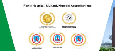 Fortis Hospital, Mulund, Mumbai4.6
4.6
-
 Fortis Hospital, Mohali4.6
4.6
-
 Fortis Hospital, Kalyan, Mumbai4.5
4.5
-
 Fortis Hospital, Anandpur, Kolkata4.1
4.1
-
 Fortis Escorts Hospital, Jaipur4.4
4.4
-
 Fortis Escorts Hospital, Amritsar4.1
4.1
More Procedures
View all
Barium enema
-
Bone Density Test
-
Breast MRI
-
Carotid Angioplasty and Stenting
-
Carotid Ultrasound
-
Chest X-rays
-
Computed Tomography (CT scan)
-
Discogram
-
Heart Scan (Coronary Calcium Scan)
-
Magnetic Resonance Imaging (MRI)
-
Mammogram
-
Needle biopsy
-
Positron Emission Tomography (PET) Scan
-
Rradio-imaging
-
Ultrasound

Keep track of your appointments, get updates & more!